Home
/ Pelvic Anatomy Labeled : Surgical Anatomy Of The Female Pelvis By Laparoscopy : The pelvis's frame is made up of the bones of the pelvis, which connect the axial skeleton to the femurs, and therefore acts in weight bearing of the upper body.
Pelvic Anatomy Labeled : Surgical Anatomy Of The Female Pelvis By Laparoscopy : The pelvis's frame is made up of the bones of the pelvis, which connect the axial skeleton to the femurs, and therefore acts in weight bearing of the upper body.
Pelvic Anatomy Labeled : Surgical Anatomy Of The Female Pelvis By Laparoscopy : The pelvis's frame is made up of the bones of the pelvis, which connect the axial skeleton to the femurs, and therefore acts in weight bearing of the upper body.. Posters ship separate from stickers. The pelvis's frame is made up of the bones of the pelvis, which connect the axial skeleton to the femurs, and therefore acts in weight bearing of the upper body. The pelvis is the lower portion of the trunk, located between the abdomen and the lower limbs. There are four articulations within the pelvis: A pelvic floor anatomy poster made by sandyspines, perfect for your office!
The pelvic bones are smaller and narrower. Corpus cavernosum of the penis. Digital illustration of pelvic girdle in colour background. It is inferior to the pelvic diaphragm. Pelvis, human skeleton, female pelvic bone anatomy, hip, 3d artwork, bones labeled anatomy view, white background.
Module 5 Pelvis Imaging from www.hitachihealthcare.com Posters ship separate from stickers. Minimalist and simple logo, flat style, modern icon and symbol. Anatomical structures of the abdomen and pelvis are visible as interactive labeled images. The poster is 16x20 or18x24 and printed on a lightly glossy heavy weight poster paper. The pelvis is the lower portion of the trunk, located between the abdomen and the lower limbs. When you are taking anatomy and physiology you will be required to know the anatomical structure locations of the pelvis. The floor of the pelvis is made up of the muscles of the pelvis, which support its. The labeled structures are (excluding the correct side):
There are four articulations within the pelvis:
Minimalist and simple logo, flat style, modern icon and symbol. Anatomical structures of the abdomen and pelvis are visible as interactive labeled images. The hip bone is an irregularly shaped bone, also known as the pelvic girdle. This quiz is unlabeled so it will test your knowledge on how to identify these structural locations (iliac crest, ischial spine, acetabulum, superior ramus of pubis, posterior superior/inferior iliac spine, lessier. Third part of the duodenum. In an adult, the innominate bones consist of the fused ilium, ischium, and pubis (figure 1). It is inferior to the pelvic diaphragm. The pelvis's frame is made up of the bones of the pelvis, which connect the axial skeleton to the femurs, and therefore acts in weight bearing of the upper body. There are four articulations within the pelvis: Almencla female pelvis and pelvic muscle models human anatomy bone medical 1:1. The female pelvis is slightly different from the male pelvis. There is a printable worksheet available for download here so you can take the quiz with pen and paper. Corpus cavernosum of the penis.
Click on the tags below to find other quizzes on the same subject. See female pelvic anatomy stock video clips. Corpus cavernosum of the penis. Third part of the duodenum. Tendinous intersection within the rectus abdominis muscle.
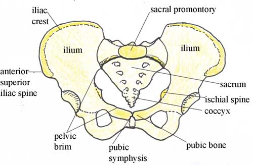
The Pelvis Human Anatomy And Physiology Lab Bsb 141 from s3-us-west-2.amazonaws.com Anatomy of female pelvic area facebook twitter linkedin pinterest print fertility and reproductive health pelvic floor disorders fertility, pregnancy and childbirth women's health. Digital illustration of pelvic girdle in colour background. In an adult, the innominate bones consist of the fused ilium, ischium, and pubis (figure 1). The bony pelvis consists of the two hip bones (also known as innominate or pelvic bones), the sacrum and the coccyx. The pelvis (plural pelves or pelvises) is either the lower part of the trunk of the human body between the abdomen and the thighs (sometimes also called pelvic region of the trunk) or the skeleton embedded in it (sometimes also called bony pelvis, or pelvic skeleton). Pelvic girdle anatomy labeled : Über 7 millionen englischsprachige bücher. The hip bone is an irregularly shaped bone, also known as the pelvic girdle.
The pelvis is the lower portion of the trunk, located between the abdomen and the lower limbs.
Diagram of pelvic bone, female pelvic bone anatomy diagram, male pelvic bone diagram, pubic bone diagram during pregnancy, what is the pelvic bone diagram, bone. The pelvic region is the area between the trunk — or main body — and the lower extremities, or legs. See female pelvic anatomy stock video clips. Third part of the duodenum. It is inferior to the pelvic diaphragm. Magnetic resonance imaging or mri of the female pelvis offers a unique display of the pelvic anatomy, including a woman's ovaries, uterus, and fallopian tubes. Digital illustration of pelvic girdle in colour background. This quiz is unlabeled so it will test your knowledge on how to identify these structural locations (iliac crest, ischial spine, acetabulum, superior ramus of pubis, posterior superior/inferior iliac spine, lessier. 3.7 out of 5 stars 4. Anatomy of the pelvis bony anatomy. This is an online quiz called label the pelvis. Regarding the surface anatomy, the perineal area is the region between the thighs, extending from the pubic symphysis anteriorly to the gluteal folds posteriorly. There is a printable worksheet available for download here so you can take the quiz with pen and paper.
Anatomy of female pelvic area facebook twitter linkedin pinterest print fertility and reproductive health pelvic floor disorders fertility, pregnancy and childbirth women's health. The pelvic bones are smaller and narrower. The poster is 16x20 or18x24 and printed on a lightly glossy heavy weight poster paper. Über 7 millionen englischsprachige bücher. The pelvis's frame is made up of the bones of the pelvis, which connect the axial skeleton to the femurs, and therefore acts in weight bearing of the upper body.
Antenatal Care Module 6 Anatomy Of The Female Pelvis And Fetal Skull View As Single Page from www.open.edu The pelvic region is the area between the trunk — or main body — and the lower extremities, or legs. The bony pelvic girdle consists of the innominate bones bilaterally, and the sacrum and coccyx posteriorly. It consists of three bones; The male pelvis is different from a female's. Mdct of the abdomen and pelvis The pelvis (plural pelves or pelvises) is either the lower part of the trunk of the human body between the abdomen and the thighs (sometimes also called pelvic region of the trunk) or the skeleton embedded in it (sometimes also called bony pelvis, or pelvic skeleton). Regarding the surface anatomy, the perineal area is the region between the thighs, extending from the pubic symphysis anteriorly to the gluteal folds posteriorly. Axis scientific flexible female pelvis anatomy model with l4 and l5 vertebrae.
Ascending colon superior mesenteric vein superior mesenteric artery gonadal vessels linea semilunaris abdominal aorta linea alba inferior vena cava inferior mesenteric artery infe.
The labeled structures are (excluding the correct side): There are four articulations within the pelvis: The bony pelvic girdle consists of the innominate bones bilaterally, and the sacrum and coccyx posteriorly. Mdct of the abdomen and pelvis Pelvis (hip) anatomy quiz for anatomy and physiology! Anatomical structures of the abdomen and pelvis are visible as interactive labeled images. Reproduction system pelvis female woman reproductive system pelvic floor women female bladder and urethra female pelvic floor pelvis woman pelvic floor health woman incontinence pelvis muscles. Pelvis, human skeleton, female pelvic bone anatomy, hip, 3d artwork, bones labeled anatomy view, white background. Click on the tags below to find other quizzes on the same subject. 3.7 out of 5 stars 4. The lining of the uterus. Axis scientific flexible female pelvis anatomy model with l4 and l5 vertebrae. Minimalist and simple logo, flat style, modern icon and symbol.
Ascending colon superior mesenteric vein superior mesenteric artery gonadal vessels linea semilunaris abdominal aorta linea alba inferior vena cava inferior mesenteric artery infe pelvic anatomy. Pelvis (hip) anatomy quiz for anatomy and physiology!